
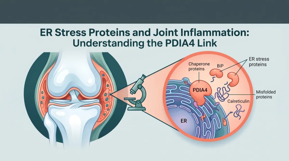
ER Stress Proteins and Joint Inflammation: The PDIA4 Discovery
Table of Contents
Introduction: A New Piece of the Osteoarthritis Puzzle
Myth 1: "Scientists have found the cause of osteoarthritis."
Myth 2: "A PDIA4-blocking treatment is on the way."
Myth 3: "Stress causes arthritis."
Myth 4: "Osteoarthritis is just mechanical wear."
Myth 5: "These results prove ER stress proteins drive human joint inflammation."
Introduction: A New Piece of the Osteoarthritis Puzzle
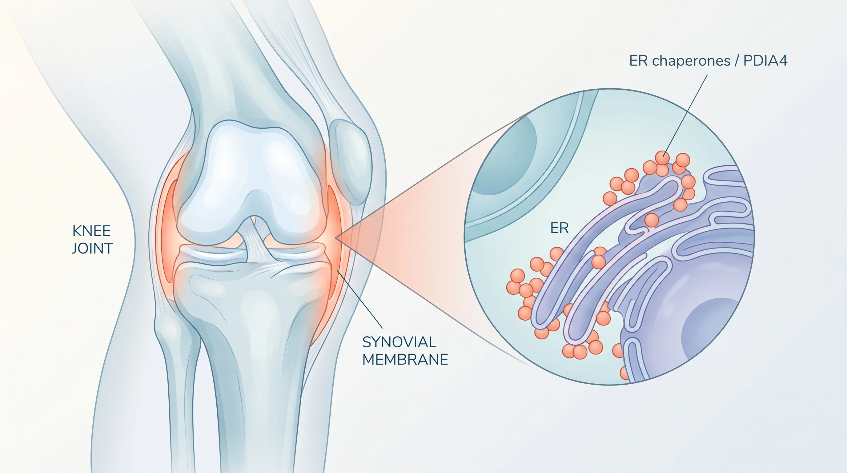
Osteoarthritis was long described as simple "wear and tear." We now know that the synovial membrane lining the joint often becomes inflamed (synovitis), but why that inflammation persists instead of resolving has remained unclear.
A study published in August 2026 in Cell Death & Differentiation by researchers at the University of Liège, the GIGA Institute and CHU de Liège (Belgium) offers a partial answer. Examining ER stress proteins and joint inflammation in human synovial tissue, the team found that one of these proteins, PDIA4, appears to actively help joint-lining cells multiply and spread, two hallmarks of chronic synovitis.
The endoplasmic reticulum (ER) is the cell's protein factory. Under sustained pressure, misfolded proteins accumulate and the ER launches the unfolded protein response, producing more chaperones that assist folding. This is basic laboratory research on human tissue and isolated cells: it identifies a possible therapeutic target, not a treatment.
The Study at a Glance
Design: Laboratory research; no patients were treated.
Scope: 11 ER stress proteins (including BiP, HYOU1, MANF, PDIA4 and calreticulin) mapped in synovial tissue from osteoarthritis, rheumatoid arthritis and calcium pyrophosphate arthropathy.
Samples: 24 biopsies for standard staining, 3 for imaging mass cytometry, 25 osteoarthritis biopsies for confirmation, cultured joint-lining cells from knee-replacement patients, and a supporting mouse model.
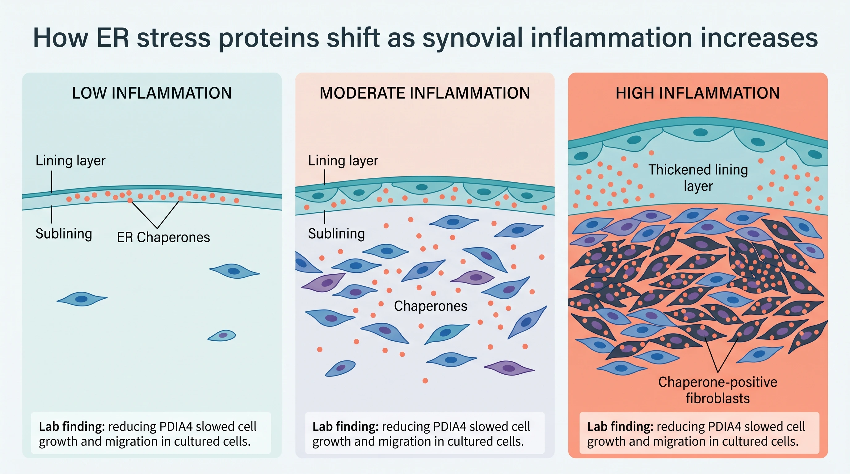
Key observations: As inflammation intensified, ER stress proteins spread from the thin lining layer into the deeper sublining. In highly inflamed samples, the share of deeper CD34⁺ fibroblasts carrying PDIA4 rose from under 10% to roughly a third. Silencing PDIA4 in cultured cells with siRNA significantly reduced their TGF-β-driven growth and migration without affecting viability, and PDIA4 appeared to be regulated mainly through the ATF6 pathway.
Myths vs. Clinical Reality
Myth 1: "Scientists have found the cause of osteoarthritis."
Clinical Reality: Osteoarthritis is a whole-joint disease shaped by age, load, injury, weight, metabolism and genetics. This study addresses one component: synovial inflammation. As specialists in the field, we observe that its value lies precisely in that focus.
Myth 2: "A PDIA4-blocking treatment is on the way."
Clinical Reality: No drug targeting PDIA4 in the joint exists or has been tested in people. siRNA is a laboratory tool, not a medicine, and the authors note that PDIA4's role in living joints remains to be established. Because cells throughout the body rely on this enzyme family, selective joint targeting would also be a considerable challenge.
Myth 3: "Stress causes arthritis."
Clinical Reality: ER stress is a molecular overload of the cell's folding machinery, not emotional stress.
Myth 4: "Osteoarthritis is just mechanical wear."
Clinical Reality: Synovitis is common in osteoarthritis and has been associated with symptoms and progression. These findings reinforce the disease's active biological dimension.
Myth 5: "These results prove ER stress proteins drive human joint inflammation."
Clinical Reality: Tissue data show correlation, not causation; the detailed imaging used only three biopsies; cell cultures were mixed populations; human samples came largely from older women while the mice were young males; and prior research suggested the same protein (ERp72) may be protective in another arthritis model. As specialists, we observe that these are the normal boundaries of early mechanistic research.
When Should You See a Specialist About Joint Pain?
While this research points toward future possibilities, the practical question for most readers is simpler: when does joint discomfort warrant a medical evaluation? The following criteria reflect widely accepted clinical guidance.
Consider scheduling an evaluation with a board-certified rheumatologist, orthopedic specialist or sports medicine physician if you experience:
Joint pain lasting more than a few weeks that does not improve with rest and reasonable activity changes.
Visible swelling, warmth or a sense of fullness in a joint, which can indicate synovial inflammation or fluid accumulation.
Morning stiffness that lasts longer than about 30 minutes, or stiffness that improves with movement. Prolonged morning stiffness can suggest an inflammatory arthritis, such as rheumatoid arthritis, which requires a different approach from osteoarthritis.
Pain in several joints at once, especially symmetrically (both hands, both wrists, both knees).
Loss of function: difficulty climbing stairs, gripping objects, walking usual distances or sleeping because of joint pain.
Locking, catching or giving way of a joint, which may point to a mechanical problem such as a meniscal tear.
Seek urgent medical care if:
A joint becomes suddenly red, hot and very painful, especially with fever or chills. This can indicate infection or an acute crystal arthritis and needs prompt assessment.
Joint pain follows a significant injury with inability to bear weight or obvious deformity.
What a specialist evaluation typically involves:
A detailed history and physical examination of the affected joints.
Imaging when appropriate (X-ray, ultrasound or MRI), which can reveal structural changes and, in the case of ultrasound or MRI, signs of synovitis.
Blood tests when an inflammatory or autoimmune arthritis is suspected.
In some cases, analysis of joint fluid.
For osteoarthritis specifically, current evidence-based management continues to center on exercise therapy, physical therapy, weight management when relevant, and appropriate pain control, tailored to the individual by a qualified clinician. Emerging laboratory findings like the one discussed here do not change that standard of care.
Conclusion
The Liège study helps explain why osteoarthritis inflammation can become self-sustaining: ER stress proteins follow inflammation deeper into the joint lining, and PDIA4 appears to participate in the growth and movement of the cells that remodel it. Yet the evidence remains preclinical. PDIA4 is a credible research target, not a therapeutic promise, and persistent joint symptoms deserve proper clinical evaluation today with proven tools.
This content reflects the professional opinion of Dr. Aleksandr Orlov of the PrecisionBio Clinic team, provided when consulted on the research discussed, under the editorial oversight of Carlos López, CEO of PrecisionBio Clinic. It is for educational purposes only and does not constitute medical advice, diagnosis or treatment. PDIA4 is a potential target under investigation, not an available treatment. For joint pain, swelling or stiffness, please consult your primary care physician or a board-certified specialist.